Technical Data Precision optical, mechanical and electrical integration instruments, integration of the three-dimensional motion system, auto-focus shooting control system, optical light imaging system, image display processing systems and auxiliary systems, using non-contact mode, you can make a quick auto-calibration, automatic shooting of corneal endothelial cell images and measure the thickness of the cornea, meanwhile, embedded in a rapid and efficient functional software for analysis of corneal endothelial cell images, can make a detailed analysis of the captured images in a timely manner, so as to give help and support to the quick check and analysis of cornea status Main performance index
On-screen photography magnification: 165X±10%;
Photography fissure width: 0.25mm±0.025mm;
Accuracy of corneal thickness measurement: ±0.025mm(>0.6mm),±0.01mm(≤0.6mm);
Shooting measuring point: center of the cornea, nasal, temporal side, upper side and lower side of the other seven solid point of view
Weight: 25Kg
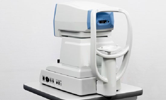
Size: 360mm×380mm×450mm Features: Dual CCD focus, you can also observe the eye and endothelial Non-contact measurement system to make rapid measurements more secure, convenient, simultaneous display of corneal thickness Built-in workstations, integrating multiple analysis and measurement tools Provides automatic / semi-automatic / manual image acquisition mode 3D AF Color LCD display, touch screen input 7:00 fixation standard design available corneal center, nasal, temporal side, upper side and lower side of the other image Support for external video duplicator Supports USB data export functions
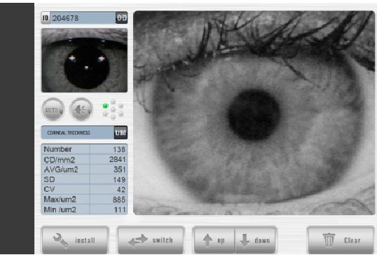
Software features: Can display the number of endothelial density, standard deviation, coefficient of variation, average / maximum / minimum Crochet automatic, manual Crochet, coloring, amplification, automatic analysis and other functions According to cell size and cell number of sides on the statistical classification of cells
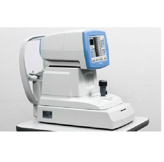
Corneal endothelial cell counter HE7000
Technical Data Precision optical, mechanical and electrical integration instruments, integration of the three-dimensional motion system, auto-focus shooting control system, optical light imaging system, image display processing systems and auxiliary systems, using non-contact mode, you can make a quick auto-calibration, automatic shooting of corneal endothelial cell images and measure the thickness of the cornea, meanwhile, embedded in a rapid and efficient functional software for analysis of corneal endothelial cell images, can make a detailed analysis of the captured images in a timely manner, so as to give help and support to the quick check and analysis of cornea status Main performance index
On-screen photography magnification: 165X±10%;
Photography fissure width: 0.25mm±0.025mm;
Accuracy of corneal thickness measurement: ±0.025mm(>0.6mm),±0.01mm(≤0.6mm);
Shooting measuring point: center of the cornea, nasal, temporal side, upper side and lower side of the other seven solid point of view
Weight: 25Kg
Size: 360mm×380mm×450mm Features: Dual CCD focus, you can also observe the eye and endothelial Non-contact measurement system to make rapid measurements more secure, convenient, simultaneous display of corneal thickness Built-in workstations, integrating multiple analysis and measurement tools Provides automatic / semi-automatic / manual image acquisition mode 3D AF Color LCD display, touch screen input 7:00 fixation standard design available corneal center, nasal, temporal side, upper side and lower side of the other image Support for external video duplicator Supports USB data export functions
Software features: Can display the number of endothelial density, standard deviation, coefficient of variation, average / maximum / minimum Crochet automatic, manual Crochet, coloring, amplification, automatic analysis and other functions According to cell size and cell number of sides on the statistical classification of cells