Welcome To Nanjing Redsun Optical Co., Ltd.
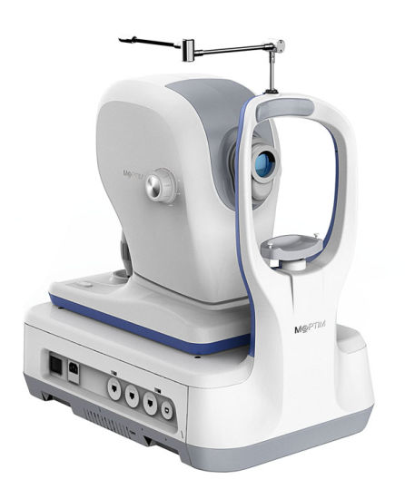
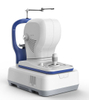
MOcean4000
redsun
9018500000
| Quantity: | |
|---|---|

Optical coherence Tomography
(MOcean 4000)
Technical Data
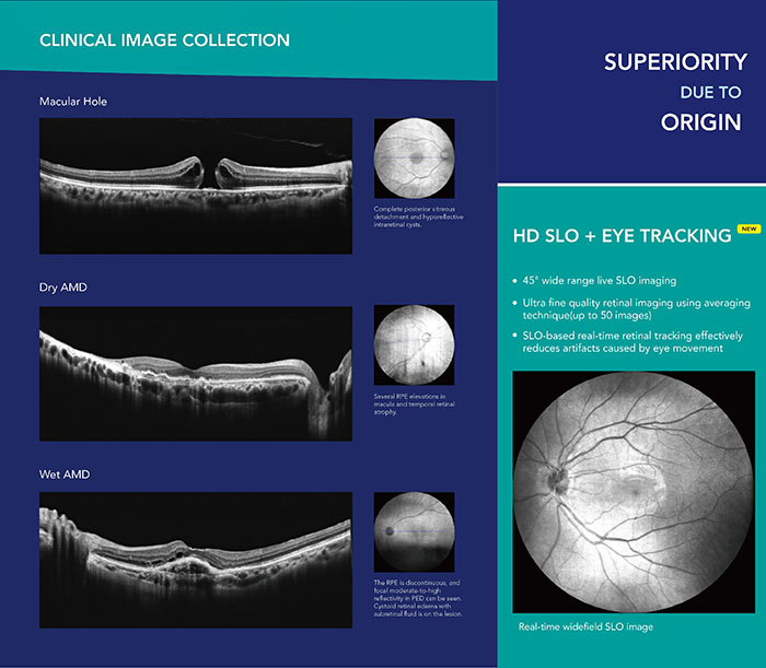
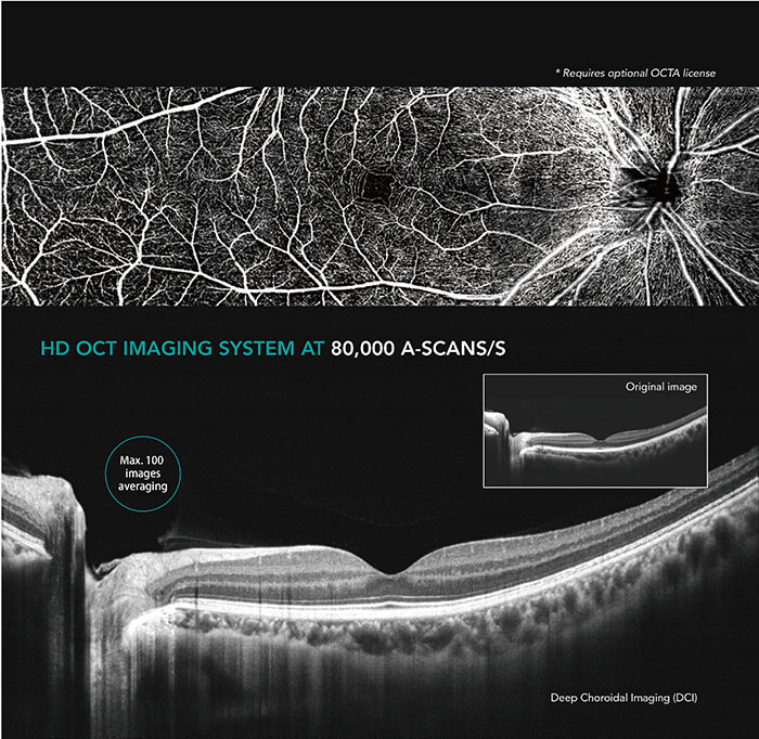
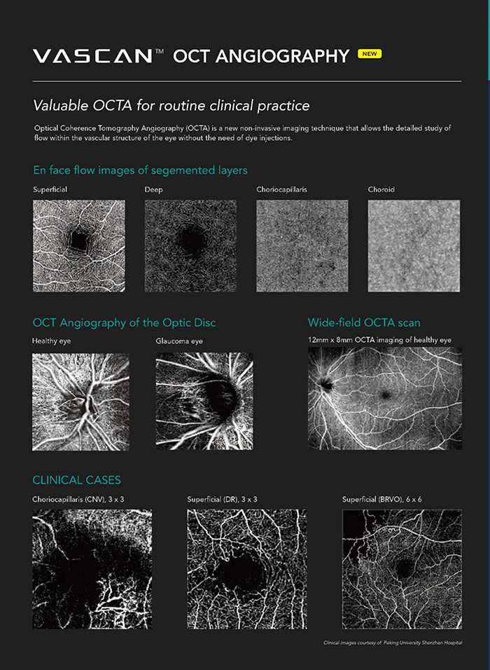
Superior OCT image quality with up to 100 times averaging
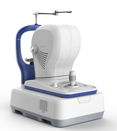
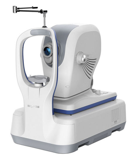
Mocean 4000
HD SLO + retinal tracking
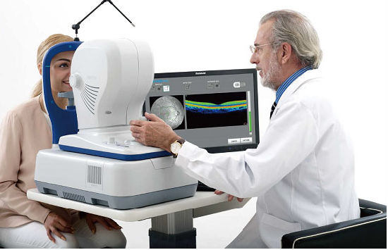
The key advantage of Mocean® 4000 system is the simultaneous acquisition of cross-sectional OCT imaging and 45 degrees fundus imaging based on Scanning Laser Ophthalmoscope (SLO). It gives you an overview of the retina so you can easily locate the lesion area before acquisition. Moreover, the system captures up to 50 SLO fundus images within one second in order to generate an HD fundus imaging with enhanced signal-to-noise ratio.
To minimize the artifacts caused by eye drift and micro saccades, Mocean® 4000 uses SLO-based eye tracker. It performs 100 times tracking per second with 10 microns tracking accuracy and more than 95% success rate, which gives you more confidence in practice.
Mocean 4000
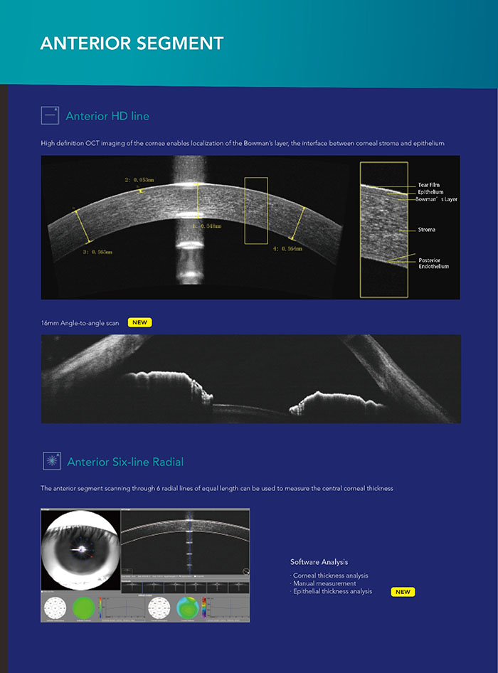
16mm angle-to-angle scan
Mocean 4000
Comprehensive analysis of retina, glaucoma and cornea
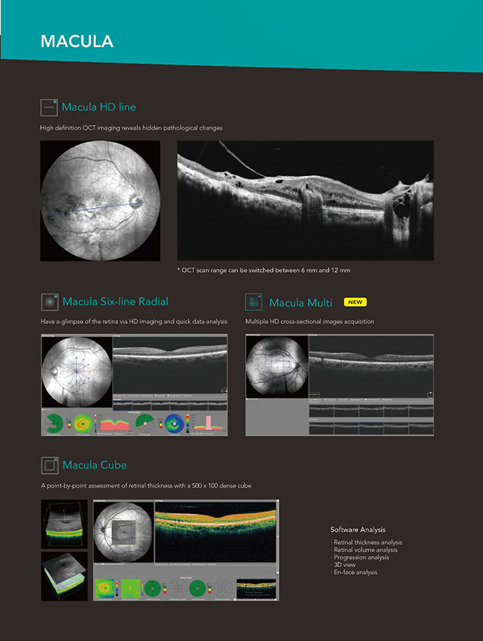
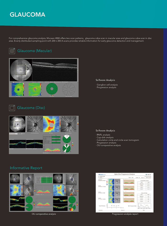
The Mocean® 4000 system provides 8 scan patterns to help you improve diagnostic efficiency:
Retina (HD line, six-radial lines, multi, cube)
Glaucoma (macular cube, disc cube)
Cornea (HD line, six-radial lines)
The software analysis features are always and up-to-date and free for upgrade (excluding OCTA module).
SPECIFICATIONS
OCT IMAGING | |
Methodology | Spectral Domain Oct |
Optical Source | Super luminescent diode(SLD),840nm |
Scan speed | 8000 A-Scan/s |
Axial Resolution(optical) | 5 microns(optical)2.7 microns(digital) |
Tranverse resolution | 15 microns(optional),3 microns9digital) |
A-Scan depth | 3MM |
Diopter range | -20D to +20D |
Scan patterns | Macular :HD line scan(6mm / 12mm),3D scan(6mm*6mm), 6-Line radial scan , multi(X-Y:5*5) Disc:3D scan(6mm*6mm) Anterior:HD Line scan: (6/16mm), 6-line radial scan |
FUNDUS IMAGING | |
Methodology | Line scanning opthalmoscopoy(LSLO) |
Minmum pupil diameter | 3.0mm |
Field of view | 45 degrees |
VASCAN TM OCTA MODULE | |
Scanning volume/area | VASCAN Advance VASCAN Essential 3mm *3mm 256*256 A scan 3mm*3mm 256*256 A scan 6mm*6mm 360*360 A scan 12mm*8mm 540*360 A scan 8mm*8mm 360*360 A scan 12mm*8mm 540*360 A scan |
Algorithm | C-OMAG C-OMAG |
Segmentation options | Encoded , Vitreousretina Intrerface(VRI),Deepfcial Retinal,Avascular, Choriocapillaris, Choriod ,Custom |
Quantitative analysis | Yes Not available |
ELECTRICAL AND PHYSICAL | |
Weight | 30.5kg |
Dimension | 532mm(l)*360mm(W)*540MM(H) |
Source voltage | AC 100-240V |
Frequency | 50Hz-60Hz |
Power input | 90VA |
SPECIFICATIONS SUBJECT TO CHANGED WITHOUT NOTICE | |

Optical coherence Tomography
(MOcean 4000)
Technical Data
Superior OCT image quality with up to 100 times averaging
Mocean 4000
HD SLO + retinal tracking
The key advantage of Mocean® 4000 system is the simultaneous acquisition of cross-sectional OCT imaging and 45 degrees fundus imaging based on Scanning Laser Ophthalmoscope (SLO). It gives you an overview of the retina so you can easily locate the lesion area before acquisition. Moreover, the system captures up to 50 SLO fundus images within one second in order to generate an HD fundus imaging with enhanced signal-to-noise ratio.
To minimize the artifacts caused by eye drift and micro saccades, Mocean® 4000 uses SLO-based eye tracker. It performs 100 times tracking per second with 10 microns tracking accuracy and more than 95% success rate, which gives you more confidence in practice.
Mocean 4000
16mm angle-to-angle scan
Mocean 4000
Comprehensive analysis of retina, glaucoma and cornea
The Mocean® 4000 system provides 8 scan patterns to help you improve diagnostic efficiency:
Retina (HD line, six-radial lines, multi, cube)
Glaucoma (macular cube, disc cube)
Cornea (HD line, six-radial lines)
The software analysis features are always and up-to-date and free for upgrade (excluding OCTA module).
SPECIFICATIONS
OCT IMAGING | |
Methodology | Spectral Domain Oct |
Optical Source | Super luminescent diode(SLD),840nm |
Scan speed | 8000 A-Scan/s |
Axial Resolution(optical) | 5 microns(optical)2.7 microns(digital) |
Tranverse resolution | 15 microns(optional),3 microns9digital) |
A-Scan depth | 3MM |
Diopter range | -20D to +20D |
Scan patterns | Macular :HD line scan(6mm / 12mm),3D scan(6mm*6mm), 6-Line radial scan , multi(X-Y:5*5) Disc:3D scan(6mm*6mm) Anterior:HD Line scan: (6/16mm), 6-line radial scan |
FUNDUS IMAGING | |
Methodology | Line scanning opthalmoscopoy(LSLO) |
Minmum pupil diameter | 3.0mm |
Field of view | 45 degrees |
VASCAN TM OCTA MODULE | |
Scanning volume/area | VASCAN Advance VASCAN Essential 3mm *3mm 256*256 A scan 3mm*3mm 256*256 A scan 6mm*6mm 360*360 A scan 12mm*8mm 540*360 A scan 8mm*8mm 360*360 A scan 12mm*8mm 540*360 A scan |
Algorithm | C-OMAG C-OMAG |
Segmentation options | Encoded , Vitreousretina Intrerface(VRI),Deepfcial Retinal,Avascular, Choriocapillaris, Choriod ,Custom |
Quantitative analysis | Yes Not available |
ELECTRICAL AND PHYSICAL | |
Weight | 30.5kg |
Dimension | 532mm(l)*360mm(W)*540MM(H) |
Source voltage | AC 100-240V |
Frequency | 50Hz-60Hz |
Power input | 90VA |
SPECIFICATIONS SUBJECT TO CHANGED WITHOUT NOTICE | |